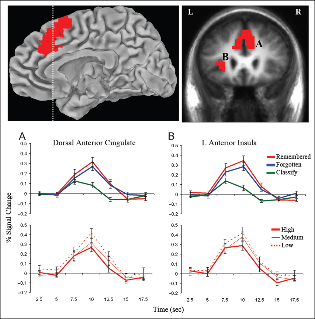
Figure 3.
Activity in DACC (A) and left anterior insula (B) increased during search and was modulated by memory strength. Statistical activation maps show the conjunction of regions with greater activity during remembered and forgotten recall trials than classify trials and increasing activity from the high- to medium- to low-study recall conditions (p < .05, corrected for multiple comparisons). Clusters are overlaid on the right medial pial surface of the Talairach and Tournoux N27 average brain and a coronal cross-section (indicated with dashed line) of the mean anatomical image of all participants. Impulse–response plots display the time course of the percent signal change (± SE) in these clusters for the remembered, forgotten, and classify trials and high-, medium-, and low-study recall conditions.

