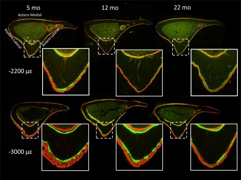
Figure 2.
Dynamic histomorphometry images of tibiae (right) from a range of adult mice subjected to tibial compression at –2200 με and –3000 με peak periosteal strain. Lamellar double-labeling was evident in all ages and both loading magnitudes, but loading at –3000 µε also induced woven bone formation.

