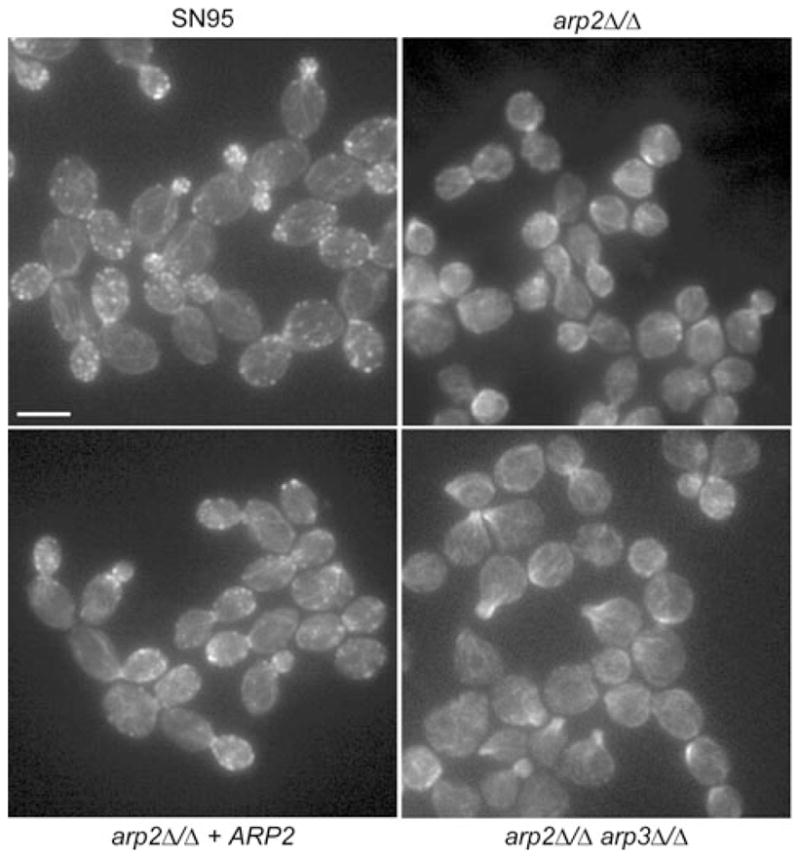
Fig. 3.

Assaying actin cytoskeleton structures in Arp2/3 complex mutants. Rhodamine/phalloidin staining was used to visualize the actin cytoskeleton in logarithmically growing yeast cells. In WT (SN95) cells, actin patches localize to sites of polarized growth in most phases of the cell cycle. Independent of the cell cycle, distinctive actin patch structures were not observed in arp2Δ/Δ or arp2Δ/Δarp3Δ/Δ cells. Actin cables seemed unaffected in mutant cells. Bar = 5 μm.
