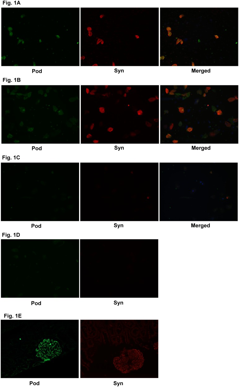
Figure 1. Images of Urinary Podocytes.
A) Representative immunofluorescent images of urinary podocytes in high risk patients with PE stained with podocin (pod), synaptopodin (syn) and colocalized (merged). B) Representative immunofluorescent images of urinary podocytes in high risk patients without PE stained with podocin (pod), synaptopodin (syn) and colocalized (merged). C): Representative immunofluorescent images of urinary podocytes in healthy pregnant control patients stained with podocin (pod), synaptopodin (syn) and colocalized (merged). D): Negative control in absence of primary antibody E) Positive control of podocin (pod) and synaptodin (syn) on normal kidney tissue.

