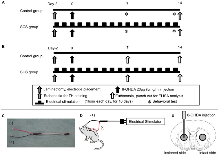
Figure 1. Time course and SCS electrode, and the brain region punched out for protein assay.
(A) Scheme showing overall experimental design. (B) Scheme showing experimental design for protein assay. (C) Photograph showing SCS electrode used in this study (diameter: 2 mm; wire length: 60 mm). (D) Scheme showing a rat during stimulation. (E) Brain tissue (diameter: 3 mm showing gray circle), corresponding to the striatum, was punched out from both the lesioned and the intact side.

