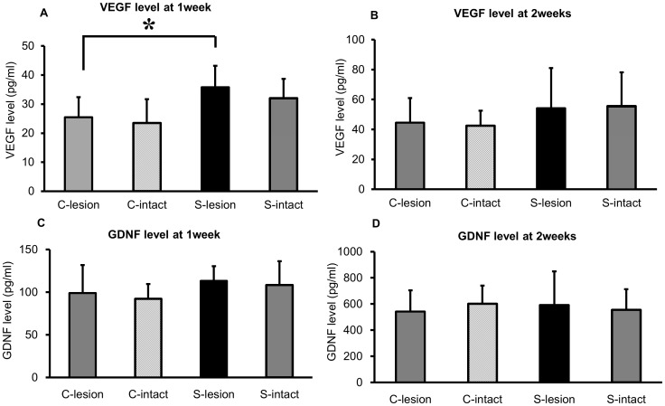
Figure 5. Results of ELISA analysis for VEGF and GDNF.
(A, B) In the lesioned striatum, VEGF was significantly increased by SCS at 1 week after 6-OHDA lesion (*p<0.05). At 2 weeks after 6-OHDA lesion, VEGF level in the lesioned striatum also appeared elevated, but did not reach statistical significance. (C, D) GDNF in the striatum of both sides was not significantly increased by SCS at 1 and 2 weeks after 6-OHDA lesion. (C-lesion: control group lesioned side Striatum; C-intact: control group intact side Striatum; S-lesion: 50 Hz SCS group lesioned side Striatum; S-intact: 50 Hz SCS group intact side Striatum, n = 10, respectively).

