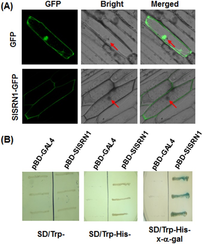
Figure 2. Subcellular localization and transactivation activity of SlSRN1.
A. Subcellular localization of SlSRN1 when transiently expressed in onion epidermal cells. Onion epidermal cells were transiently transformed with either control GFP vector (upper) or SlSRN1-GFP construct (lower) by particle bombardment. The subcellular localization of the SlSRN1-GFP fusion protein and GFP alone were viewed at 24 hr after bombardment under a confocal laser microscopy in dark field for green fluorescence (left), in white field for the morphology of the cell (middle), and in combination (right), respectively. Red arrows indicate the nucleuses of the onion epidermal cells. B. Transactivation activity of SlSRN1 in yeast. Yeasts carrying pBD-SlSRN1 or pBD empty vector (as a negative control) were streaked on the SD/Trp− plates (left) or SD/Trp−His− plates (middle) for 3 days at 28°C. The x-α-gal was added to the SD/Trp−His− plates and kept at 28°C for 6 hr (right).

