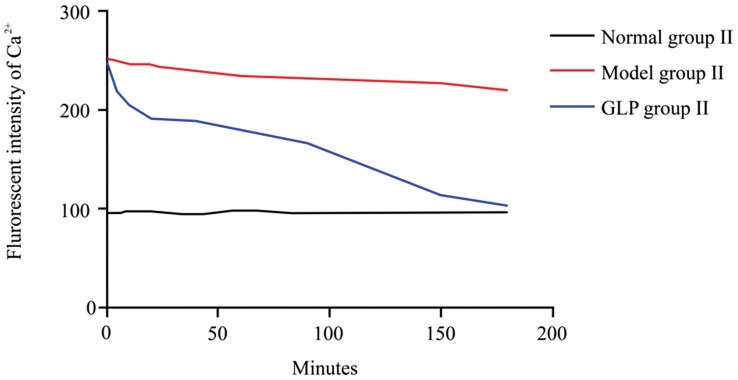
Figure 2. Changes in fluorescence intensity of Ca2+ in neurons during the treatment, n = 4, thirty neurons were observed in each replicates.
There is no change in fluorescence intensity of Ca2+ in neurons in the Control group II during a 3 hour period. Fluorescence intensity of Ca2+ in neurons was highest in neurons cultured in Mg2+ ion free medium for 3 hours prior to replacement with normal maintaining medium i.e. Model group II; there is no significant decrease (P>0.05) between the zero time point (0 hour) and 3 hours following the normal medium replacement. However, following GLP treatment, there is a gradual decrease in Fluorescence intensity of Ca2+ in neurons, with levels almost returning to normal after 3 hours. Tukey’s test following one way ANOVA was used to perform statistical analysis.

