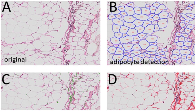
Figure 1. Adipose immunohistochemistry for cell size and fibrosis.
Adipose tissue was stained for elastin, as described in Methods. A. Elastin stain, illustrating the outline of the adipocytes, elastin (black stain) and areas of fibrosis (pink). B. Identification of adipocytes using the image analysis software, and the assignment of adipocyte area to the cells. C. Enhancement of image to bring out elastin (green). D. Enhancement of image to bring out collagens (red).

