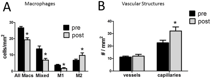
Figure 3. Adipose macrophage polarity and vascularity following pioglitazone treatment.

A. Macrophages were characterized as M1, M2, or mixed, as described in Methods, pre and post pioglitazone treatment. B. Adipose blood vessels were characterized as capillaries, or vessels, based on the absence/presence of ASMA staining of a vessel wall (see Methods) and counted in adipose sections pre and post pioglitazone. *p<0.05 vs pre pioglitazone.
