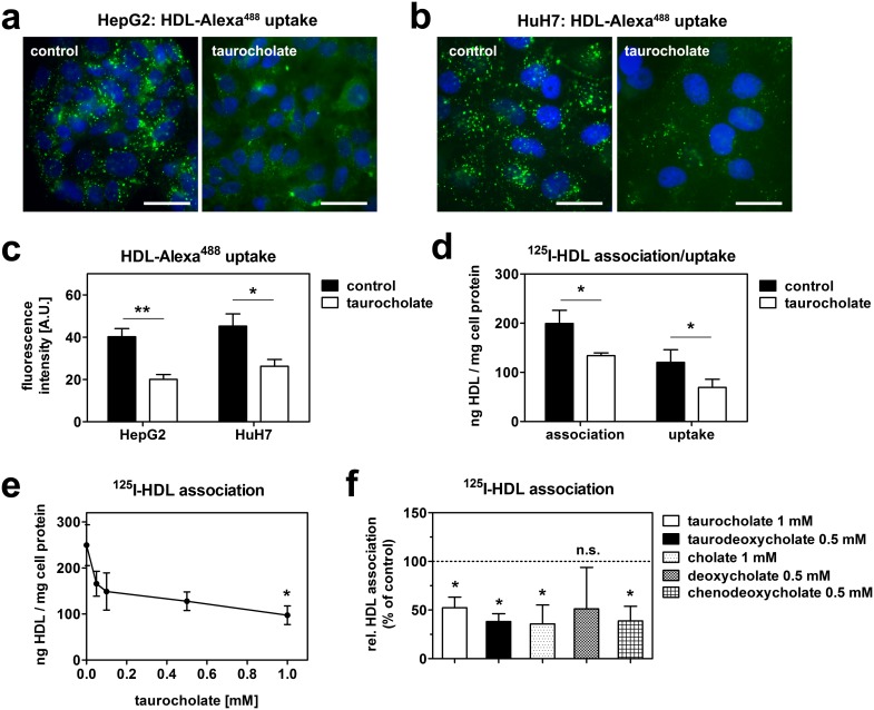
Figure 1. Bile acids reduce HDL endocytosis.
HepG2 (a) and HuH7 (b) cells were incubated with 50 µg/ml HDL-Alexa488 with or without 1 mM taurocholate at 37°C for 1 hour. Cells were fixed, counterstained with DAPI and imaged. Green: HDL; blue: nucleus; bar = 10 µm. Representative images of 3 independent experiments are shown. (c) Quantification of fluorescence intensities of (a) and (b). (d) HepG2 cells were incubated in media containing 20 µg/ml 125I-HDL with or without 1 mM taurocholate at 37°C for 1 hour. Uptake was determined after displacing cell surface bound HDL by a 100-fold excess at 4°C for 1 hour (n = 3). (e) Cells were incubated with 20 µg/ml 125I-HDL with the indicated concentrations of taurocholate for 1 hour (n = 3). (f) Cells were incubated with 20 µg/ml 125I-HDL together with different bile acids for 1 hour (n = 3). Of note taurodeoxycholate, deoxycholate and chenodeoxycholate were cytotoxic at 1 mM and were therefore used at 0.5 mM.

