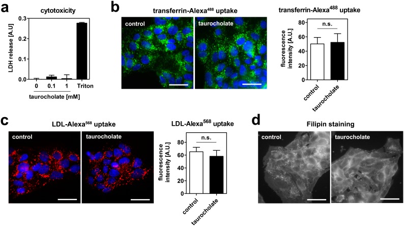
Figure 2. Taurocholate neither exerts cytotoxic effects, nor inhibits transferrin or LDL endocytosis in HepG2 cells.
(a) Cells were incubated with the indicated concentrations of taurocholate for 1 hour. No release of LDH into the cell culture supernatant was detected. 0.1% Triton-X100 was used as a positive control. (b) Cells were incubated with 20 µg/ml transferrin-Alexa488 (b) or 50 µg/ml LDL-Alexa568 (c) with or without 1 mM taurocholate at 37°C for 1 hour. Cells were fixed, counterstained with DAPI and imaged. Green: transferrin; red: LDL; blue: nucleus; bar = 10 µm. Neither transferrin nor LDL uptake were altered. Quantifications of fluorescent signals are depicted next to the images. (d) Cells were incubated with or without 1 mM taurocholate for 1 hour. Cells were fixed, stained with Filipin and imaged. Bar = 10 µm. Representative images of 3 independent experiments are shown.

