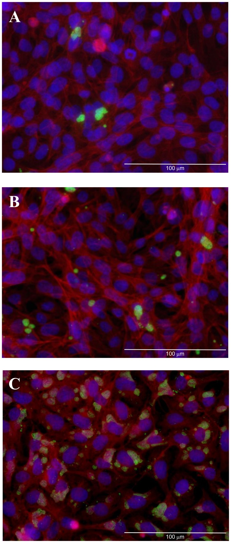
Figure 1. Fluorescent micrographs demonstrating the infectivity of W. chondrophila at an apparent MOI of 0.1 (A), 1 (B) and 10 (C) at 24 h post-infection.
W. chondrophila inclusions are labelled green using an anti-Waddlia rabbit polyclonal antisera and FITC anti-rabbit secondary antibody, host cell nuclei are stained in blue (Dapi) and host actin-filaments labelled with Phalloidin-Atto 550 (red). The scale bars correspond to 100 µm.

