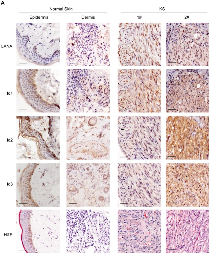
Figure 4. Ids were aberrantly expressed in KS tissues.
Expression of Id1, Id2, Id3, and LANA were detected in 10 KS tissues and 5 normal skin tissues by immunohistochemistry. There were weak to modest staining signals of Id1, Id2 and Id3 only in the basal cells of epidermis and around the hair follicle of dermis in normal skin tissues. There was no LANA staining in any normal skin tissues. In contrast, there were strong staining signals of Id1, Id2, Id3 and LANA in the spindle tumor cells in KS lesions. Representative images of the immunohistochemistry staining were shown.

