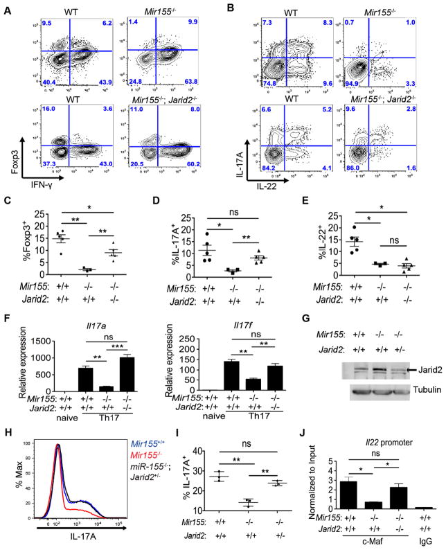
Figure 7. Epistasis between Jarid2 and miR-155 in Treg and Th17 cells.
(A,B) FACS analysis of WT, Mir155−/− or double deficient Mir155−/−; Jarid2fl/fl; CD4-cre (Mir155−/−; Jarid2−/−) cells from siLP of infected mixed BM chimeras to enumerate CD4+TCRβ+CD44+ cells that are IFN-γ or Foxp3 positive (A), or IL-17A and/or IL-22 positive (B), 8 days after T. gondii infection. (C–E) Indicated percentages of CD4+TCRβ+CD44+ WT, Mir155−/− or Mir155−/−; Jarid2−/− cells from the siLP of infected mixed BM chimeras. (F) Relative expression of Il17a and Il17f mRNA was determined by RT-qPCR in WT, Mir155−/− or Mir155−/−; Jarid2−/− Th17 cultures (− ecogenous IL-1β). (G) Western blot analysis of Jarid2 and tubulin protein in WT, Mir155−/− and Mir155−/−; Jarid2+/− Th17 cells. (H) Histogram depicts intracellular IL-17A expression in WT, Mir155−/− and Mir155−/−; Jarid2+/− Th17 cultures (− IL-1β). (I) The corresponding percentages of CD4+CD44+IL-17A+ T cells. (J) c-MAF and IgG ChIP-qPCR on the Il22 promoter in WT, Mir155−/− and Mir155−/−; Jarid2−/− Th17 cultures normalized to input. Statistical significance determined using unpaired Student’s t test (* p<0.05, ** p<0.01, and *** p<0.001; ns denotes not significant). ns denotes not significant.

