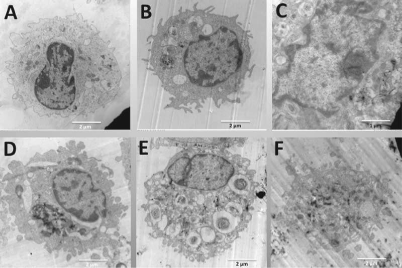
Figure 7.
TEM representative images of individual C57BL/6 alveolar macrophages (AM) incubated with the five 60_100 long multi-walled carbon nanotubes (MWCNT). All particle exposures were at 50 μg/ml in 500 μl at 106 cells/ml AM in suspension culture. After 1 h all AM samples were collected, fixed, and processed as described in Methods section for transmission electron microscopy analyses. (A) Control AM with no particle exposure at 7000×. (B) AM exposed to FA04 at 10,000×. (C) AM exposed to FA08B at 15,000×. (D) AM exposed to FA10B at 9000×. (E) AM exposed to FA17 at 7000×. (F) AM exposed to FA21 at 8000×. Lysosomal formation is evident in B and D (low nickel particles), whereas MWCNT free in the cytoplasm are evident in C, E, and F (high nickel particles).

