Fig. 7.
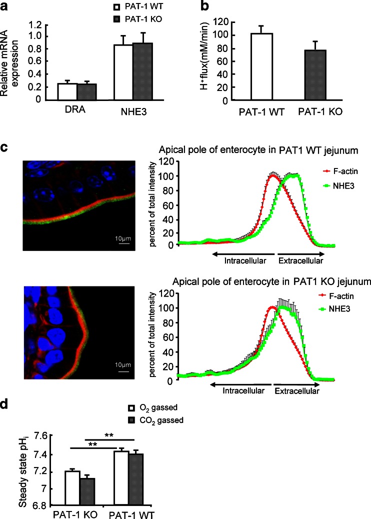
Absence of jejunal PAT-1 expression results in low enterocyte pHi, but not in an altered NHE3 expression, membrane localization or functional activity. a DRA and NHE3 mRNA expression levels in PAT-1 KO and WT jejunum. b Acid-activated NHE3 transport activity was assessed as the HOE642 insensitive, S1611-sensitive Na+-dependent proton export rate in microdissected jeunal villi of PAT-1 KO and WT enterocytes, as described in “Material and methods”. No significant difference was seen between the two genotypes (b); n = 3–7, *p < 0.05. c NHE3 staining in the jejunal enterocyte brush border membrane in relation to the F-actin apical cytoskeleton (phalloidin staining), and quantitation of the relative distribution of NHE3 in % of total brush border membrane NHE3 within the analysed area, along the terminal web-microvillar axis (left panels). The peak of the F-actin intensity indicates the microvillar cleft/terminal web zone [28]. It is evident that the majority of NHE3 is located more towards the lumen, i.e., in the microvilli, both in WT and PAT-1 KO jejunum. Eight different cellular regions (different cells, different villi) were studied from three pairs of mice. d Steady-state pHi in PAT-1 KO and WT enterocytes in the absence or presence of CO2/HCO3 −.*p < 0.05, n = 5–7

