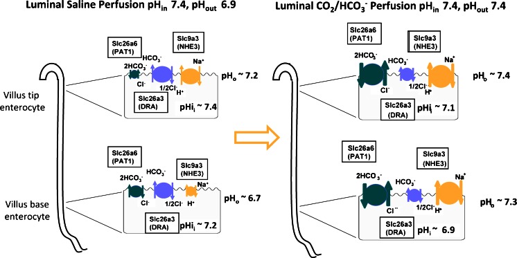
Fig. 9.
Hypothetical schematic diagram of the ion transport activities in the jejunal enterocytes at the upper and lower part of the villus, based on the (approximate) measurements of intra- and extracellular pH and the expected contributions of the different transporters to fluid absorption based on measurements in WT and KO mice. The size of the transporter symbols is indicative of presumed activity. Left panel: during perfusion of the jejunal segment with unbuffered saline, the perfusate rapidly acidifies due to CO2 diffusion from the tissue, H+ export via NHE3 and very little HCO3 − absorption via PAT-1. Some HCO3 − is likely formed near the enterocyte membrane by extracellular carbonic anhydrases. DRA activity alkalinises the perfusate. The high pHi favours DRA-mediated HCO3 − secretion, and it is likely that even PAT-1 will import Cl− in exchange for HCO3 −, at least in the acidic extracellular pH conditions at the base of the villi. The low pHo has an inhibitory effect on NHE3. Right panel: after the switch of the perfusate to a CO2/HCO3 − buffer with the same pH, the surface pHo is stable at 7.4, while CO2 enters and acidifies the enterocytes. This creates a driving force for PAT-1 mediated HCO3 − absorption; it increases the driving force for NHE3-mediated Na+ absorption, while reducing it for DRA-mediated HCO3 − secretion

