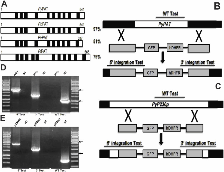
Figure 1. Structure, conservation of PAT and Targeted deletion of PAT and P230p.
(A). Schematic representation of the putative PyPAT protein organization and alignment with PAT proteins from other Plasmodium species. The transmembrane domains are shown as black boxes. Total amino acid sequence identities to PyPAT are shown to the right. (B. and C.) Schematic representation of the replacement strategy to generate Pypat(-) parasites in (B) and Pyp230p(-) parasites in (C). The endogenous PyPAT and PyP230p genomic loci are targeted with replacement fragments containing the 5′ and 3′ PyPAT UTRs and PyP230p ORFs sequences, respectively, flanking the human DHFR positive selection marker and eGFP cassettes. Diagnostic WT-specific or integration-specific test amplicons are indicated by lines. (D. and E.) PCR genotyping confirms the integration of gene-replacement construct using oligonucleotide primer combinations that can only amplify from the recombinant loci (5′ Test and 3′Test). The WT-specific PCR reaction (WT) confirms the absence of WT parasites in Pypat(-) parasites in (D) and in Pyp230p(-) parasites in (E).The arrows showing the size of DNA ladder bands of 1000 and 3000 bps, respectively.

