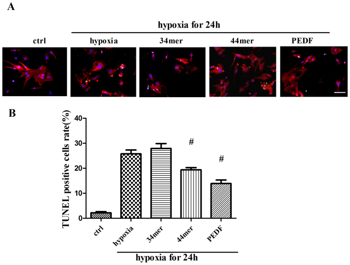
Figure 5. PEDF and 44mer protected against hypoxia-induced cell death in cultured neonatal rat myocardial cells.
(a) The purity of neonatal myocardial cells was about 95% identified by α-sa staining. TUNEL (green) and α-sa (red) staining were performed at each group (control, 24 h hypoxia with or without 34mer, 44mer, PEDF). Nucleis were stained with Hoechst (blue). Immunofluorescent images showed that TUNEL positive cells (arrow indicated) were increased after hypoxia. PEDF and 44mer protected against TUNEL positive cells rate, while 34mer had no effect. (b) Statistic analyses from immunofluorescent images. (n = 10; #P < 0.05 compare to hypoxia group). Scale bar, 20 μm. Data are represented as means ± S.E.M.

