Figure 2.

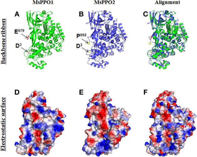
Electrostatic surfaces of MsPPO1 and MsPPO2. Crystal structures of two Manduca sexta PPOs (PDB ID code 3HHS) were aligned using the PyMOL Molecular Graphics System (http://pymol.org/). (A–C) Secondary structure (Backbone ribbons) of two M. sexta PPOs. PPO1 crystal structure (A). The N-terminus residue is D3, and C-terminus residue is E679 as indicated by arrows. PPO2 crystal structure (B). The N-terminus residue is D3, and C-terminus residue is E692 as indicated by arrows. (C). Alignment of PPO1 and PPO2 at the same angle. (D,E) The electrostatic surfaces of two M. sexta PPOs after being generated using the PyMOL software. The backbone ribbons and electrostatic surfaces of each PPO are surveyed from the same view. Red is negative, and blue is positive. MsPPO1 (D) and MsPPO2 (E) have different surface electrical charges. In (D), the circled negative area is composed of I97D98, A221D222 residues. In (E), the circled negative area is composed of N97E98, D101, S225A226S227, E229, V232, S355V356L357 residues. (F). Alignment of PPO1 and PPO2 at the same angle.
