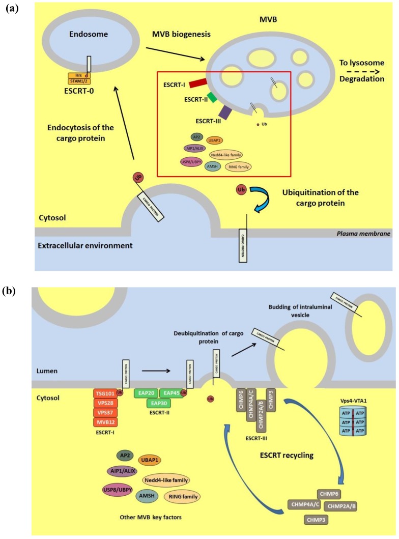
Figure 3.
(a) Schematic representation of the MVB biogenesis pathway. An enlargement of the red squared part is shown in panel b; (b) Schematic representation of the vesiculation process leading to the formation of the MVB. The sequential recruitment of the ESCRT complexes to the MVB membrane is described along with the additional factors involved in the cargo protein delivery into the organelle lumen. The extracellular environment and its equivalents are colored in light blue, while the cytoplasmic environment is colored in yellow. Details on the ESCRT proteins and on the other MVB key factors can be found in several comprehensive reviews [7,169,173,201,202].

