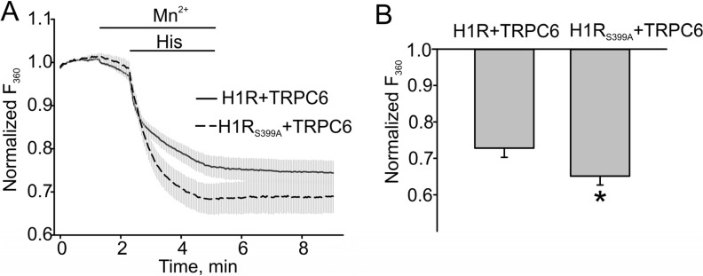
Figure 4.
The H1 receptor mutant potentiated the TRPC6 channel activity in HEK model. (A) Mn2+ (0.5 mM) influx in H1R/TRPC6 expressing HEK cells versus H1RS399A/TRPC6 expressing HEK cells, which were measured by the percent of Fura-2 quenching (Fura-2 was excited at 360 nm) at the time point of 5 min. (B) The summary data comparing the percent of Fura-2 quenching are shown. The bold and broken lines represent the averaged traces, and the vertical gray lines are the SEM values. Data in (B) are presented as mean ± S.E. Asterisk = p < 0.05.

