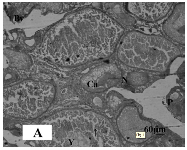
Microphotograph 1A.

Histological section of ovary of control Oreochromis niloticus showing oocyte in the first growth phase (→), cortical alveolar cells (Ca), Perinucleaur cells (P), Blood vessels (Bv), Yolk bodies (Y), and Nucleolus of oocytes. Haematoxylin and Eosin, X-bar 60 μm.
