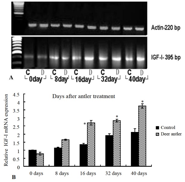
Figure 3.

Comparison between RT-PCR and real-time RT-PCR of IGF-I at indicated the days post-injury. A: RT-PCR was performed on RNA isolated from new hair occurring around wound samples. To determine relative changes of IGF-I in mRNA levels during hair and wound healing development, RT-PCR was performed using specific primers and fractionated on 1.0% agarose gel. The size of the predicted amplified product for IGF-I (395 bp) is indicated on the right. A 220 bp β-actin fragment was amplified from the same RT reaction to serve as an endogenous internal control. C = Control group; D = Deer antler group. B: Real-time RT-PCR of IGF comparative expression in the indicated days post-injury.
