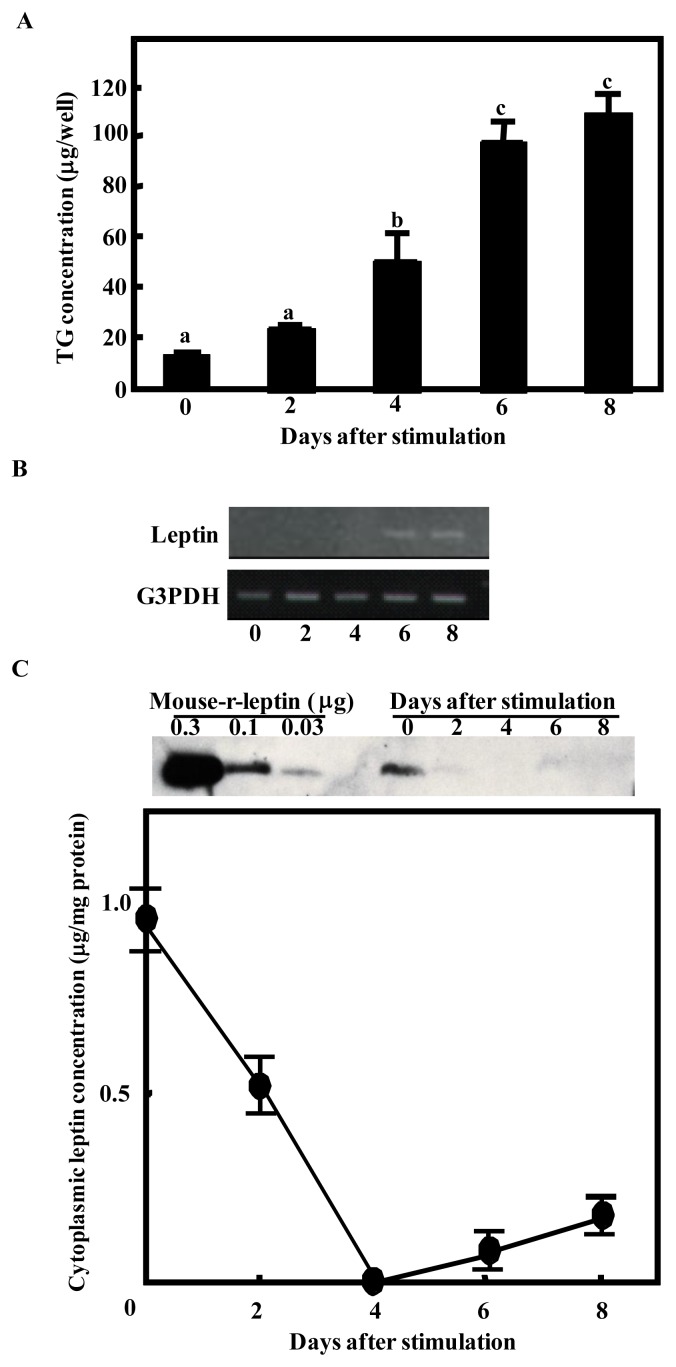
Figure 1.
Leptin mRNA expression level and cytoplasmic leptin concentration in bovine intramuscular preadipocyte cells (BIP cells) during differentiation. After reaching confluence, adipocyte differentiation was initiated by the addition of differentiation factors. (A) Triglyceride (TG) accumulation was assessed in BIP cells on the day after induction. Points with a different superscript (a, b, c) are significantly different (p<0.05) (B) RT-PCR of the expression of leptin (upper panel) and G3PDH (lower panel) mRNA in BIP cells on the day indicated after induction. The results are representative of three similar experiments. (C) Cell lysates were collected at the day indicated after induction. Proteins were separated by SDS-PAGE and analyzed by immunoblotting using a polyclonal antibody for leptin. Recombinant murine leptin (Murine r-leptin) protein was included at 1.0, 0.3, and 0.1 μg per well as a positive control. The intensities of the bands were analyzed using NIH Image software and murine leptin was used as a standard to quantify bovine leptin. Y-axis indicates that leptin contents per 1 mg cytoplasmic proteins. The results represent the mean±SEM for three independent determinations. This is representative of three similar experiments.

