Figure 2.
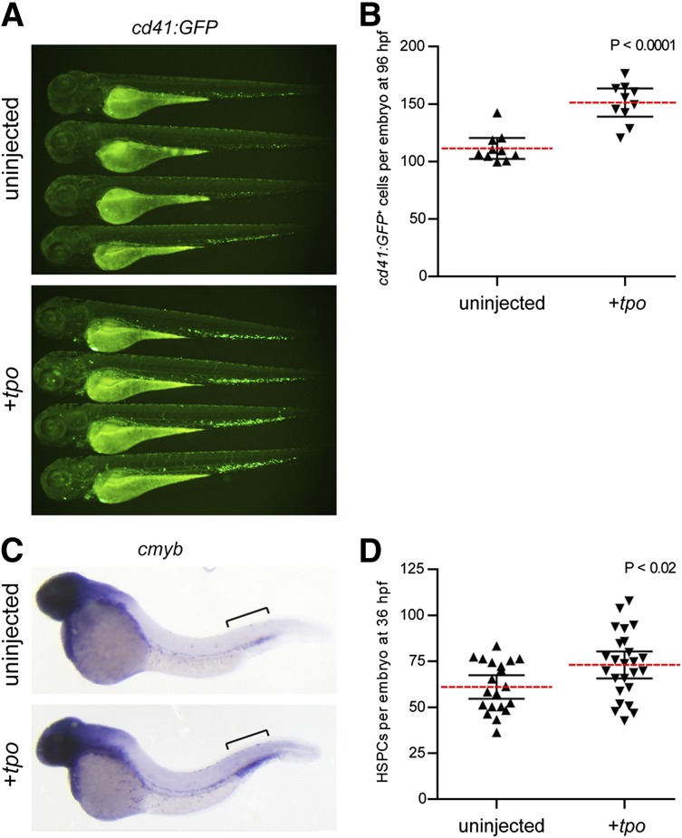
tpo expands the number of thrombocytes and HSPCs in the developing zebrafish embryo. (A) cd41:GFP+ cells in 72 hpf zebrafish embryos uninjected (top) or injected with tpo mRNA (bottom). (B) Numbers of cd41:GFP+ cells quantitated from 96 hpf embryos. Embryos were analyzed using flow cytometry, and the number of cd41:GFP+ cells was calculated based on the frequency of these cells multiplied by the total cell number per embryo. Each data point represents 5 embryos pooled together before digestion and flow cytometry. (C) cmyb+ HSPCs present in the caudal hematopoietic tissue (brackets) at 36 hpf in embryos that were uninjected (top) or injected with tpo mRNA (bottom). (D) Numbers of cmyb+ cells along the dorsal aorta and caudal hematopoietic tissue (brackets) region at 36 hpf quantitated from 2 independent experiments. Dashed red lines in B and D represent the mean with 95% confidence interval (black error bars).

