
Figure 3.
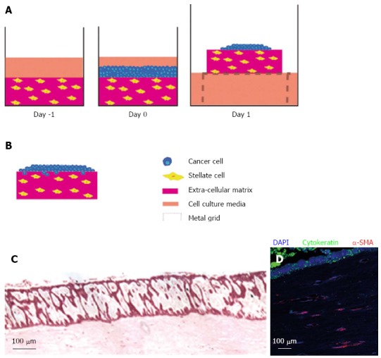
Raised organotypic model of pancreatic ductal adenocarcinoma with embedded pancreatic stellate cells. A: Extracellular matrix gels containing stellate cells were polymerised in 24-well plates before cancer cells were seeded on top and allowed to attach. These gels were then raised onto metal grids and fed from below creating a chemotactic gradient. Cells were cultured for up to 14 d; B: Illustration of cancer cells in a raised model containing stellate cells showing proliferation and invasion into the gel; C: HE section of a raised gel containing stellate cells with cancer cells seeded on top; D: Immunofluorscence in the same gel showing strong cytokeratin expression in the cancer cells and α-smooth muscle actin (α-SMA) expression in the embedded stellate cells.