
Figure 1.
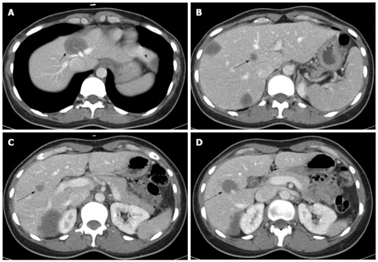
Abdominal computed tomography revealed a total of 7 lesions located in both lobes. A: The tumor of segment 4 (short arrow) hinders the possibility of right hemihepatectomy, because it invades the roots of the middle and left hepatic veins (arrowhead indicates the tumor of segment 3); B-D: The 3 deep-seated tumors in the right lobe (long arrows) hinder the possibility of left hemihepatectomy.