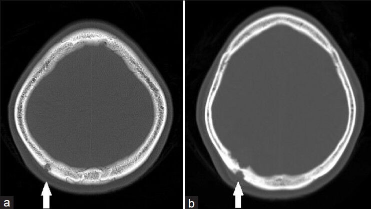
Figure 1.

(a) Initial CT brain bone window axial cuts showing lesion extending from the inner table to the outer table of the skull vault with fairly well-defined margin, in keeping with sinus pericranii with no other lytic abnormality seen in the skull vault and (b) progression of the lesion 2 years later with increased extent of bony involvement (white arrow)
