
Fig. 1.
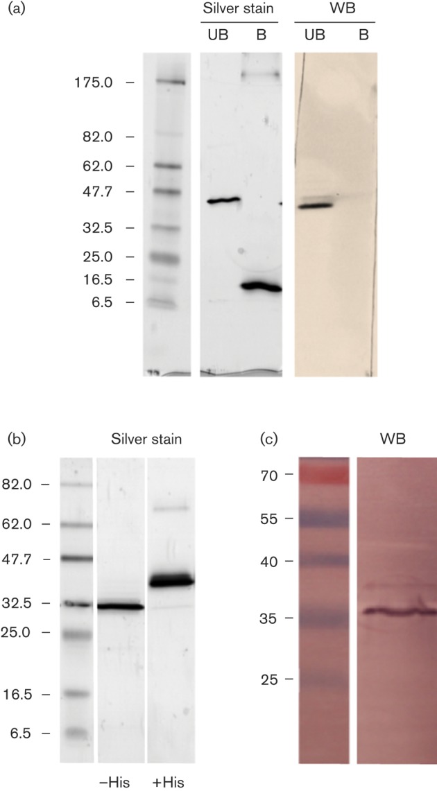
(a) SDS-PAGE separation of purified unboiled (UB) or boiled (B) KcvMA-1D with a His tag. Silver staining revealed a single band for the tetramer and for the monomer of unboiled and boiled protein, respectively. In a Western blot (WB) anti-Kcv-8D6 only recognizes the tetramer in unboiled samples. (b) KcvMA-1D with a His tag (+His) and after removing the tag (−His) in an SDS-PAGE gel stained with Coomassie blue. The tag-free channel formed a single band of ~35 kDa. (c) A Western blot of the tag-free KcvMA-1D protein using anti-Kcv-8D6 antibody detected a single band of ~35 kDa. Molecular size markers are shown on the left of blots (kDa).