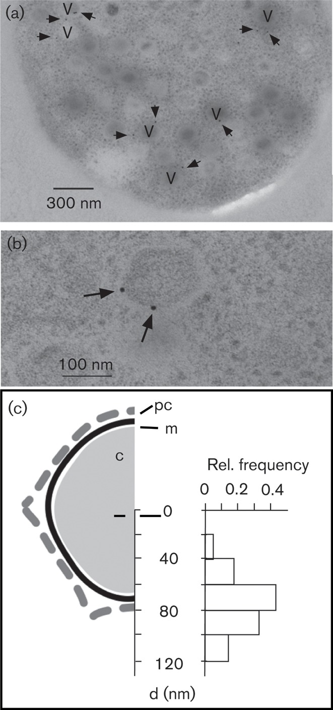
Fig. 5.

Location of the Kcv channel at the periphery of virus particles. (a) Electron micrograph of a thin section through a C. variabilis cell producing PBCV-1 particles. Virus particles are indicated with a V and gold particles are indicated by arrows. (b) Magnification of an individual virion with two peripheral gold labels. (c) Redrawing (to scale) of one half of a PBCV-1 particle (Zhang et al., 2011) showing the protein coat (pc), membrane (m) and core (c). The histogram shows the relative frequency of the distances (d) where gold particles were detected with respect to the centre of the virus particles.
