
Figure 2. Morphology of human mesenchymal stem cells and human mesenchymal cells exposed to osteogenic supplements exposed to shear stress (left to right) for 24 h.
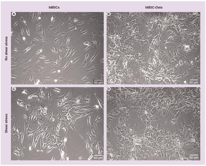
Phase contrast microscopy images of (A) hMSCs and (B) hMSC-Osts not exposed to shear stress, and (C) hMSCs and (D) hMSC-Osts exposed to shear stress. hMSCs changed their fibroblast-like, spindle-shaped morphology to a more cuboidal morphology after exposure to osteogenic supplements (A vs B). There were no obvious changes in morphology of hMSCs (A vs C) and hMSC-Osts (B vs D) after 24 h exposure to 9 dynes/cm2 steady shear stress. hMSC: Human mesenchymal cell; hMSC-Osts: Human mesenchymal cells exposed to osteogenic supplement.