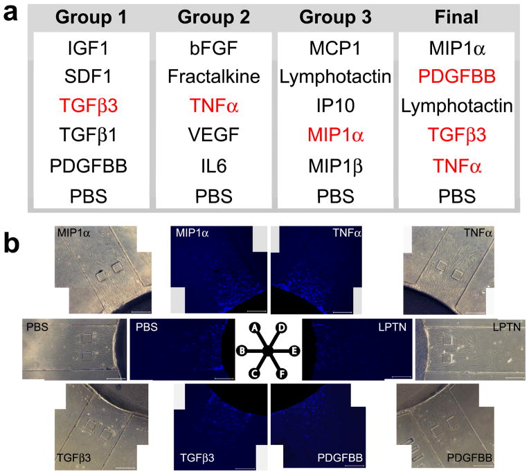
Figure 3.
Cytokine selection for cell migration assays. a: Cytokines that are crucial for arthritis were randomly allocated into 3 groups with PBS as a control for each group. The ‘top performers’ from the first three groups (in red) were selected for the ‘final’ round, which yielded the final group capable of robust cell recruitment. b: Competitive cell migration of the selected, most robust cytotactic cues. Phase contrast and DAPI stained images at 24 hrs following exposure to MIP1α, TGFβ3, TNFα, lymphotactin, PDGFbb and PBS (control). DAPI images were subsequently used to quantify the number of migrated cells, migratory distance and migration index.

