
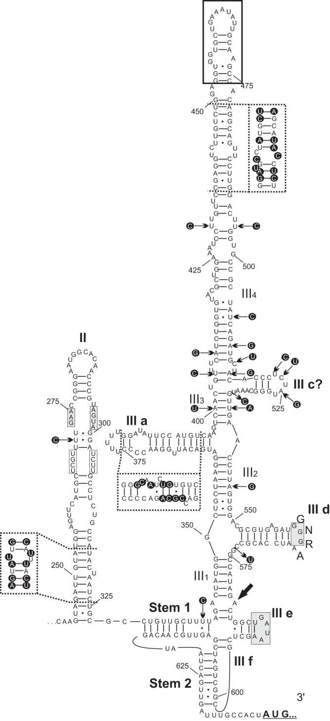
FIG 2.
Predicted two-dimensional secondary RNA structure of the 5′ UTR IRES of chicken megrivirus (GenBank accession no. KF961186) with the positions of different nucleotides (in dark circles) indicated by black arrows and within frames of dashed lines compared to the IRES of turkey/B407-THV/2011/HUN (GenBank accession no. KF961188). The main domains (II and III), helical segments (III 1 to 4), and hairpins (III a; III c to f) and stems (Stem 1 and Stem 2) similar to those of the similarly labeled structures of hepacivirus/pestivirus-like type IVB IRES are depicted. Gray boxes indicate conserved motifs of IVB IRESes (23). White boxes indicate the conserved unpaired base pairs with respect to DHAVs within the middle loop of domain II. The sequence and location of the apical “8” structure also identified in other avian picornaviruses are indicated with a black frame box (24).