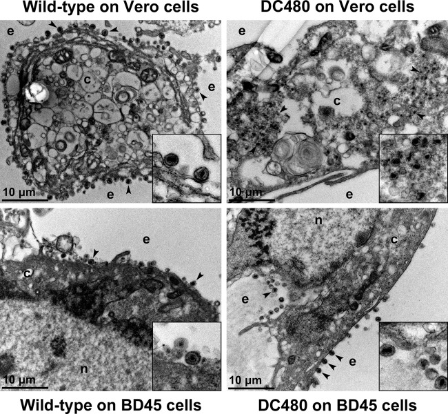
FIG 4.
Ultrastructural morphology of wild-type and mutant viruses. Electron micrographs of Vero or BD45 cells infected at an MOI of 3 with wild-type or DC480 virus and processed for electron microscopy at 18 hpi are shown. Enlarged sections of the micrographs are included as insets. The nucleus (n), cytoplasm (c), and extracellular space (e) are marked. Representative virions are marked with black arrowheads.

