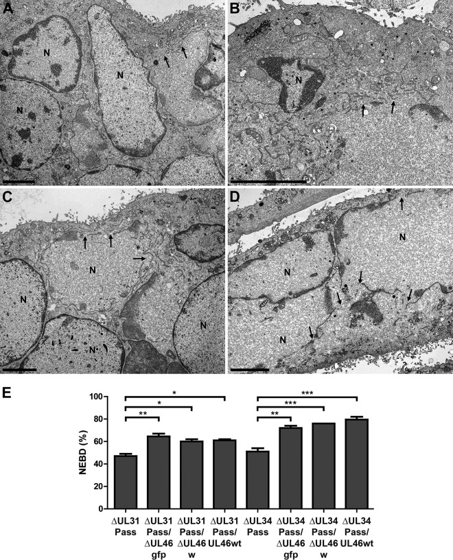
FIG 6.
Electron microscopic analysis of cells infected with UL46-deficient PrV-ΔUL31Pass and PrV-ΔUL34Pass mutants. RK13 cells were infected with PrV-ΔUL31Pass (A), PrV-ΔUL31Pass/ΔUL46gfp (B), PrV-ΔUL34Pass (C), and PrV-ΔUL34Pass/ΔUL46gfp (D) at an MOI of 1 for 14 to 18 h and subsequently processed for electron microscopy. Bars represent 4 μm, and arrows indicate NEBD. N, nucleus. (E) For quantitation of NEBD in cells infected with PrV-ΔUL31Pass, PrV-ΔUL31Pass/ΔUL46gfp, PrV-ΔUL31Pass/ΔUL46w, PrV-ΔUL31Pass/UL46wt, PrV-ΔUL34Pass, PrV-ΔUL34Pass/ΔUL46gfp, PrV-ΔUL34Pass/ΔUL46w, and PrV-ΔUL34Pass/UL46wt, approximately 70 nuclei per virus infection were assessed for ruptured or intact nuclear envelopes. Quantitation was performed twice in independent experiments, and subsequently ANOVA with a Tukey posttest was used for statistical analysis (*, P < 0.05; **, P < 0.01; ***, P < 0.001).

