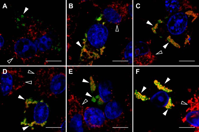
FIG 3.
NoV RdRp localizes to mitochondria, and expression of the RdRp induces mitochondrial clustering in transfected BSR-T7/5 cells. BSR-T7/5 cells were transfected with pT7-N1-HA and incubated as described in the legend to Fig. 1. At each time point, the RdRp was detected by immunostaining with anti-HA primary and FITC-labeled secondary antibodies (green). Nuclei were visualized by staining with DAPI (blue), and the morphology of mitochondrial networks was visualized by staining with MitoTracker Red CM-H2XRos (red). Each panel represents a different time point: A, 4 hpt; B, 8 hpt; C, 12 hpt; D, 16 hpt; E, 20 hpt; F, 24 hpt. Immunofluorescence confocal microscopy images with merged signals are shown; the yellow signal in the merge images indicates colocalization. Closed arrowheads indicate RdRp expression in transfected cells; open arrowheads indicate untransfected cells. Bar, 10 μm.

