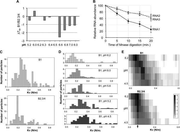
FIG 3.
B1 and B2.3/4 virions have distinct physiochemical properties. (A) Differential scanning fluorimetric analysis of the B1 and B2.3/4 virions. The difference in the TMapp of the B1 and the B2.3/4 virions is plotted. The virions were examined in phosphate buffer adjusted to the pH indicated on the horizontal axis. Each bar represents the mean of three independent measurements, and all standard errors were less than 5% of the mean. (B) The RNA in the B1 virion is more sensitive to micrococcal nuclease digestion than the RNAs in B2.3/4 virions. Virions (5 μg) at pH 7.0 were treated with 1 unit of micrococcal nuclease, and digestion was stopped by addition of 0.5 M EGTA at the indicated times. RNAs were extracted with TRIzol, precipitated, and subjected to electrophoresis in 1% agarose–TBE gels stained with ethidium bromide. The intact BMV RNAs were quantified with a Bio-Rad ImageQuant analyzer. The results are from four independent data sets. (C to E) Analysis of the B1 and B2.3/4 virions by nanoindentation using an atomic force microscope. (C) Histogram of elastic constants of B1 and B2.3/4 virions at pH 4.5. B1 virions exhibit a significantly broader distribution shifted toward stiffer elastic responses. (D) The effect of pH increase on B1 elasticity. The distribution shifts toward softer elastic responses. Note that the number of B1 virions resistant to nanoindentation decreased with increasing pH. (E) Comparison between the joint histograms of pH and elastic constants for B1 and B 2.3/4. For B2.3/4, the peak probability density at a K of 0.18 N/m is insensitive to pH changes, while for B1, it shifts gradually from a K of 0.38 N/m to 0.15 N/m.

