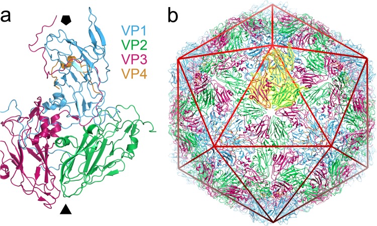
FIG 1.
Structure of equine rhinitis A virus. (a) X-ray structure of the native equine rhinitis A virus particle. A single asymmetric unit of the native ERAV virion (PDB 2WFF) is shown in diagram representation, from a view outside of the viral capsid. The viral proteins are colored blue (VP1), green (VP2), magenta (VP3), and orange (VP4), and the approximate position of the viral 5- and 3-fold axes are indicated by the solid black pentagon and triangle, respectively. The color coding of VP1 to VP4 is identical in all figures in the manuscript. (b) The intact, pT=3 capsid structure. The asymmetric unit shown in panel a is highlighted in yellow, and a icosahedron is included as a red wire mesh. The image was produced using PyMOL (www.pymol.org) and UCSF Chimera (35).

