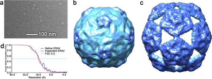
FIG 2.

Solution structure of native and expanded ERAV particles. (a) Raw cryo-electron micrograph of the ERAV sample. Two types of particle—small and large—are clearly visible in the raw data. (b) Cryo-EM structure of small ERAV particle, which corresponds to the structure of the native virion. (c) The corresponding structure of the large, expanded ERAV particle. For Fig. 2, 3, and 5, the cryo-EM-derived density for both native and expanded particles is colored with the radial color scheme described in Fig. 5. (d) Fourier shell correlation plot for both EM structures. The resolutions of the maps are 16.9 Å for the native structure and 17.4 Å for the expanded particle (both using the FSC0.5 criterion).
