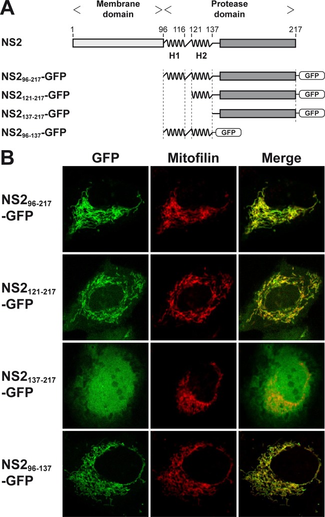
FIG 1.
Membrane association of the HCV NS2 protease domain. (A) Schematic representation of HCV NS2 and of GFP fusion constructs. The two α-helices located at the N terminus of the protease domain (comprising aa 96 to 116 [H1] and 121 to 137 [H2]) are highlighted. (B) Confocal laser scanning microscopy analyses of U-2 OS human osteosarcoma cells transiently transfected with cytomegalovirus (CMV) promoter-driven GFP fusion constructs, as illustrated in panel A. A polyclonal antibody against mitofilin (Sigma-Aldrich, St. Louis, MO) was used as a marker for mitochondria.

