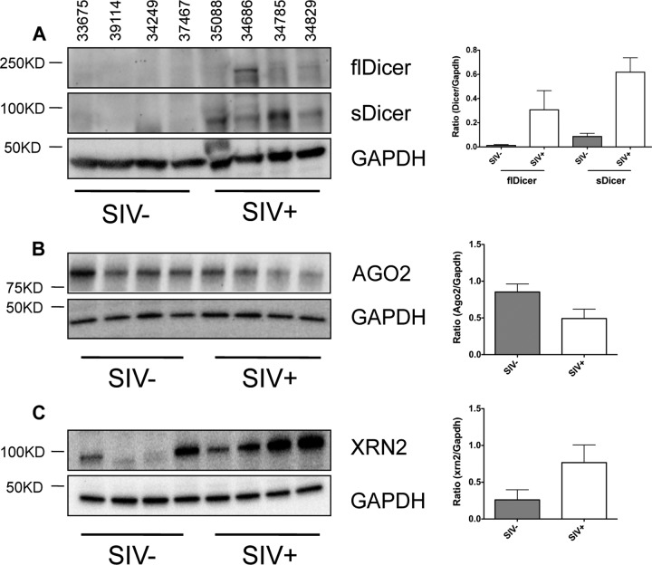
FIG 4.
Disruption of miRNA biogenesis during chronic SIV infection. Western blot analysis of DICER1 (A; left), AGO2 (B; left), and XRN2 (C; left) expression in the intestinal mucosa of SIV-infected macaques (n = 4) and SIV-negative controls (n = 4). Each lane on the blot represents a single animal, and the animal numbers are indicated above each lane of the blot. GAPDH was used as a loading control, and densitometry measurements for each protein are given as a ratio of the protein density to that of GAPDH (A to C; right). Error bars indicate the standard errors of the means.

