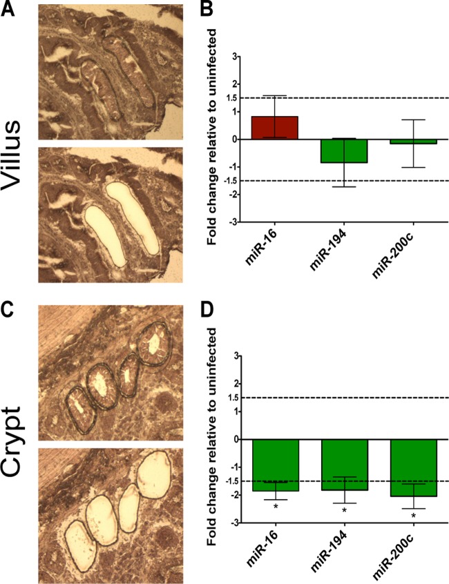
FIG 6.
Decrease in miRNA expression during chronic SIV infection is localized to the proliferative crypt epithelial region. Before and after images of villus (A) and crypt (C) epithelial cells isolated from the surrounding tissue by laser capture microdissection. (B, D) Fold changes in the expression of miR-16, -194, and -200c in the villus and crypt regions during SIV infection were determined by RT-PCR. SIV infected, n = 4; SIV negative, n = 4. Dotted lines indicate a 1.5-fold change. Statistical significance determined using Student's t test. P values of ≤0.05 are denoted by *, and error bars indicate the standard errors of the means.

