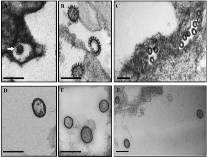
FIG 1.
Electron microscopy analysis of ISAV from eggs and ovarian fluid. (A to C) Sectioned eggs; (D) supernatant from an in vitro infection with an ISAV HPR-3 variant; (E and F) ovarian fluid. (A) The arrow shows a membranous connection between a virus particle and the membrane of the egg. (B) Magnification of three clearly distinctive viral particles inside the egg. (C) General view of numerous viral particles inside the egg. (D) Typical viral structure of an in vitro-grown ISAV. (E) Pelleted virus from ovarian fluid. (F) Direct staining of 15 μl of ovarian fluid. Scale bars, 200 nm.

