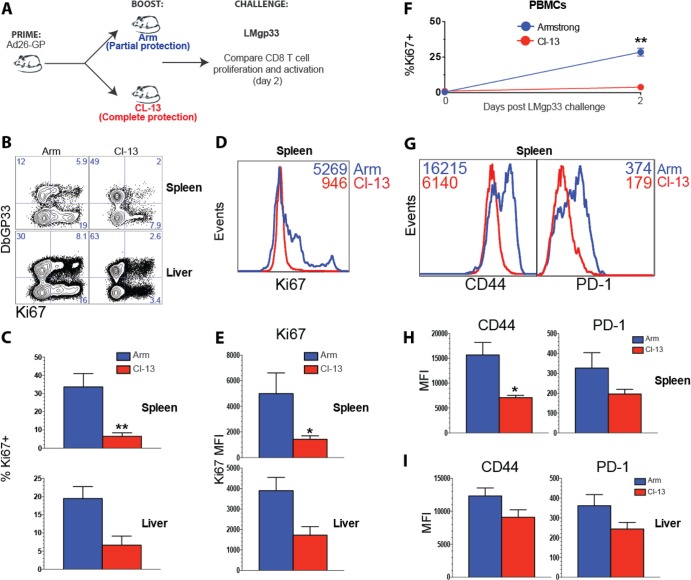
FIG 7.
Rapid immune control is associated with minimal recall expansion and modest activation of memory CD8 T cells. (A) Experimental design. Mice were primed with Ad26-GP, and after 60 days, they received an LCMV Armstrong or LCMV CL-13 boost. After >30 days, mice were challenged with LMgp33, and early anamnestic CD8 T cell responses were analyzed on day 2. (B) Representative FACS plots showing the percentages of CD8 T cells that expressed the proliferation marker Ki67. (C) Summary of the percentages of Ki67-expressing CD8 T cells. (D) Representative histograms showing Ki67 expression by splenic CD8 T cells. (E) Summary of the MFI of Ki67-expressing CD8 T cells. (F) Percentage of Ki67-expressing CD8 T cells in blood before and after Listeria challenge. (G) Representative histograms showing the mean fluorescence intensity (MFI) for CD44 and PD-1 by CD8 T cells. (H) Summary of the MFI of CD44 and PD-1 expression in spleen. (I) Summary of the MFI of CD44 and PD-1 expression in liver. Data are gated from DbGP33-specific CD8 T cells from the indicated tissues. Error bars represent SEM. Data are from two experiments (four mice per group per experiment). Values that were statistically significant are indicated by asterisks as follows: *, P = 0.05; **, P = 0.02.

