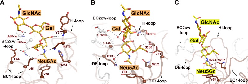
FIG 3.
Interactions between HPyV9 VP1 and sialyloligosaccharides. (A and B) Interactions between HPyV9 VP1 and 3SLN. The main chain of HPyV9 VP1 is shown in cartoon representation. One monomer is shown in chocolate color, while other monomers are shown in gray. The amino acid side chains interacting with 3SLN are shown as sticks, and the corresponding C-α atoms are shown as spheres. The backbone amide and carbonyl groups are shown only when engaging the oligosaccharide. 3SLN is shown as in Fig. 2B. Water molecules are represented as red spheres. Hydrogen bonds are shown as black dashed lines. The view in panel A was rotated by about 90° along a vertical axis to obtain the view presented in panel B. (C) Interactions between HPyV9 VP1 and 3GSLN. HPyV9 VP1 is drawn as described for panel A; the ligand 3GSLN is shown as in Fig. 2C. Residues H274 and N282, which interact specifically with the hydroxymethyl chain of Neu5Gc, are shown in stick representation.

