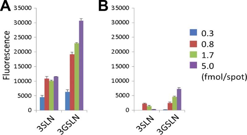
FIG 5.
Comparison of binding of the wild-type HPyV9 VP1 (A) and the mutant HPyV9 VP1 (B) to lipid-linked oligosaccharide probes (3SLN.DH and 3GSLN.DH) in microarray analyses. Both probes were arrayed at four levels (as indicated) in duplicate. Numerical scores of the binding signals are shown as the means of the fluorescence intensities of duplicate spots with error bars, which represent half of the difference between the two values. The wt and mutant HPyV9 VP1 proteins were analyzed in parallel at 150 μg/ml precomplexed with anti-His and biotin–anti-mouse IgG antibodies.

