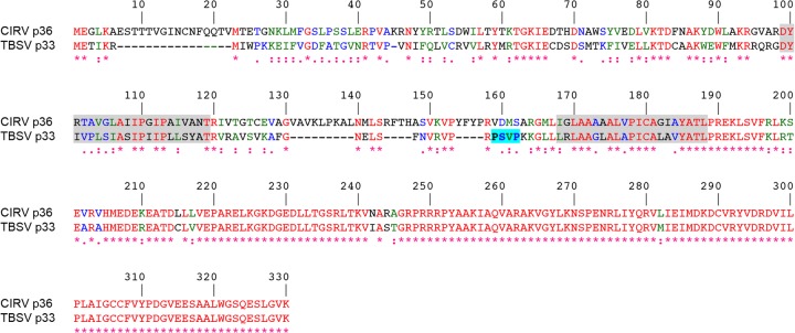
FIG 1.

Alignment of the deduced amino acid sequences of CIRV p36 and TBSV p33. Sequences were obtained from GenBank (accession numbers CAA59477.2 and NP_062898.1) and aligned using ClustalW. Identical and similar amino acids in each protein are colored red and green or blue, respectively, and indicated also with asterisks and colons or periods, respectively. The numbers represent specific amino acid residues in full-length p36 (330 residues). Putative TMDs (shown on a gray background) were determined using TOPCONS and visual inspection, and the late-domain-like motif identified in the intervening loop sequence of p33 (23) but absent in p36 is shown on a bright blue background.
