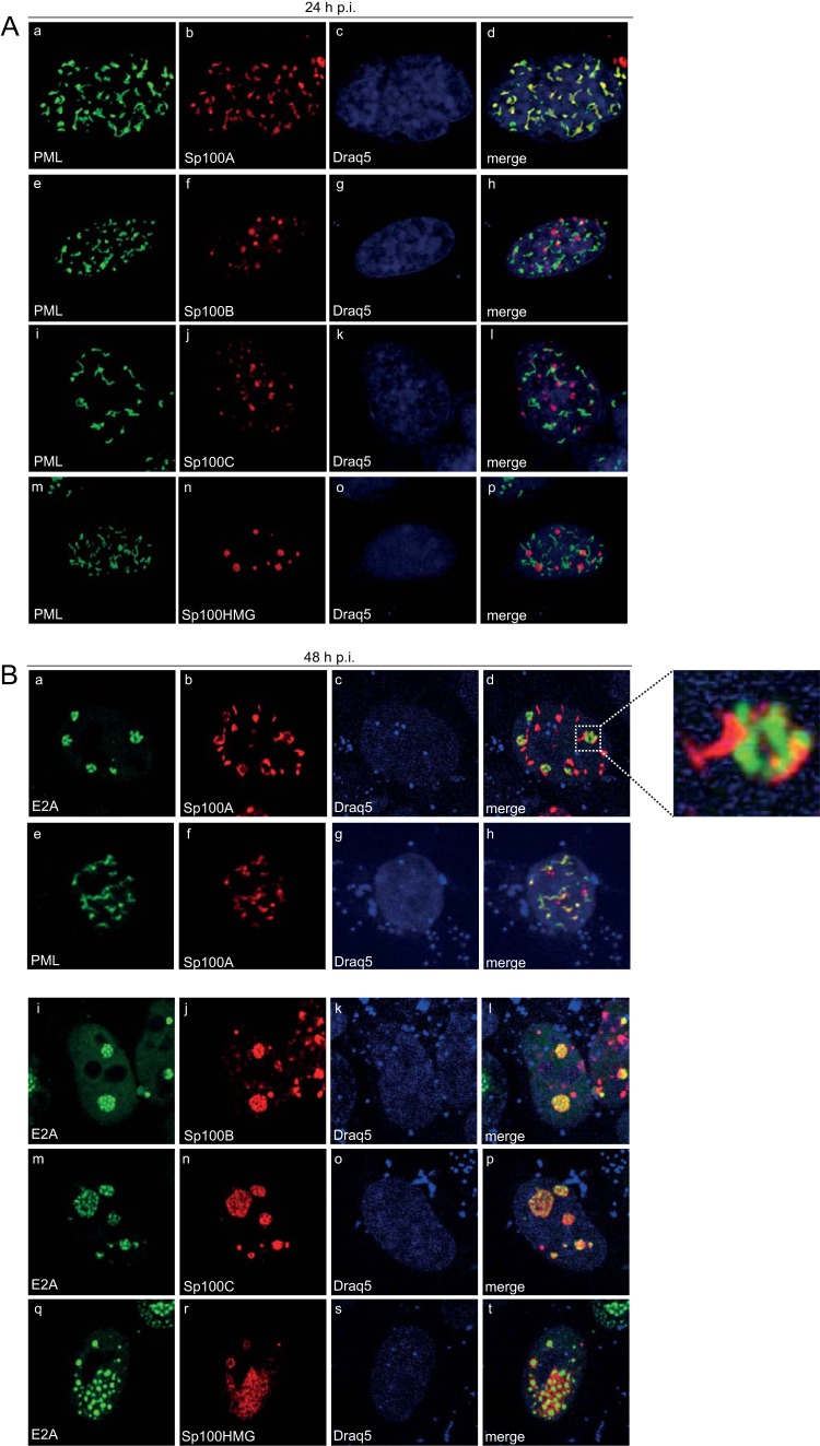
FIG 3.
Ad-dependent relocalization of Sp100B, -C, and -HMG from PML-NBs to viral replication centers. HepaRG cells were transfected with 1.5 μg of pFlag-Sp100A, -B, -C, or -HMG and superinfected with wt virus (H5pg4100) at a multiplicity of infection of 50 FFU/cell at 8 h posttransfection. (A) The cells were fixed with 4% PFA at 24 h p.i. and double labeled with MAb Flag-M2 (anti-Flag) and pAb NB 100-59787 (anti-PML). (B) The cells were fixed with 4% PFA at 48 h p.i. and double labeled with MAb Flag-M2 (anti-Flag) and pAb NB 100-59787 (anti-PML) or rabbit MAb anti-E2A. Primary Abs were detected with Cy3 (anti-Flag; red)- and Alexa 488 (anti-PML/E2A; green)-conjugated secondary Abs. The DNA-intercalating dye DRAQ5 (Biostatus) was used to stain nuclei. Anti-PML (green; A, panels a, e, i, and m, and B, panel e), anti-E2A (green; B, panels a, i, m, and q), and anti-Flag-Sp100 (red; A, panels b, f, j, and n, and B, panels b, f, j, n, and r) staining patterns representative of at least 50 analyzed cells are shown. Overlays of the single images (merge) are shown in panels d, h, l, p, and t. Magnification, ×7,600.

